loader image
Select Page

ANATOMY: COMPONENTS OF THE SPINE

The “low back” officially begins with the lumbar region of the spine directly below the cervical and thoracic regions and directly above the sacrum. The lumbar vertebrae, L1-L5, are most frequently involved in back pain because these vertebrae carry the most amount of body weight and are subject to the largest forces and stresses along the spine.

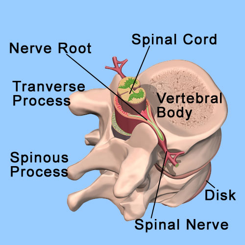
The spinal cord (bundle of nerves from the brain) ends at approximately the L1 vertebral level, where it then divides into many different nerve roots that travel to the lower body and legs. This collection of nerve roots is collectively called the “cauda equina,” which means horse’s tail and describes the continuation of the nerve roots at the end of the spinal cord.


Vertebrae
The vertebral body is a thin ring of dense cortical bone. The vertebral body is shaped like a marshmallow, thinner in the center with thicker ends. Outer cortical bone extends above and below the superior and inferior ends of the vertebrae to form rims. The superior and inferior endplates are contained within these rims of bone.
Pedicles
The pedicles are two short rounded processes that extend posterior from the lateral margin of the dorsal (towards the backside) surface of the vertebral body. They are made of thick cortical bone.
Laminae
The laminae are two flattened plates of bone extending medially from the pedicles to form the posterior wall of the vertebral foramen. The Pars Interarticularis is a special region of the lamina between the superior and inferior articular processes. A fracture or congenital anomaly of the pars may result in a spondylolisthesis (forward slippage of a vertebrae in relation to another).
Intervertebral Discs
Intervertebral discs are found between each vertebra. The discs are flat, round structures about a quarter to three quarters of an inch thick, with tough outer rings of tissue called the annulus fibrosis which contain a soft, white, jelly-like center called the nucleus pulposus. Flat, circular plates of cartilage connect to the vertebrae above and below each disc. Intervertebral discs separate the vertebrae, but they also act as shock absorbers for the spine. They compress when weight is put on them and spring back when the weight is removed.  Intervertebral discs make up about one-third of the length of the spine and constitute the largest organ in the body without its own blood supply. The discs receive their blood supply through diffusion as they soak up nutrients. The discs expand while at rest allowing them to soak up nutrient rich fluid. When this process is inhibited through repetitive movement, injury or poor posture, the discs become thinner and more prone to injury. This may be a cause of the gradual degeneration of the structure and function of the disc over time (Degenerative Disc Disease).
Facet Joints
Facet joints are paired joints (one on both sides) at every level of the spine. These joints are what allow us to bend backward and forward and twist and turn. The facet joints help support the spine, anchor ligaments and muscles, and restrict excessive spinal movement. The facet joints are part of the posterior elements of each vertebrae. Each vertebra has facet joints that connect it with the vertebrae above and the vertebrae below in the spinal column. The surfaces of the facet joints are covered with smooth cartilage that help these parts of the vertebral bodies glide smoothly over each other. Sometimes these joints become inflamed (bursitis) which results in pain.
Ligamentum Flavum
The ligamentum flavum is a strong ligament that connects the laminae of the vertebrae. The term “flavum” is used to describe the yellow appearance of this ligament in its natural state. The ligamentum flavum serves to protect the nerves and the spinal cord and stabilize the spine so that excessive motion between the vertebral bodies does not occur. It is the strongest of the spinal ligaments and often has a thinner middle section. Together with the laminae, it forms the posterior wall of the spinal canal.
Spinal Cord
The spinal cord is part of the central nervous system of the human body. It is a vital pathway that conducts electrical signals from the brain to the rest of the body through individual nerve fibers. The spinal cord is a very delicate structure that is derived from the neural tube, which eventually closes during fetal development. From this neural tube, the entire central nervous system, our brain and spinal cord, eventually develops. Up to the third month of fetal life, the spinal cord is about the same length as the canal. After the third month of development, the growth of the canal outpaces that of the cord. In an adult the lower end of the spinal cord usually ends at approximately the first lumbar vertebra, where it divides into many individual nerve roots (L1). Below the L1 Lumbar level, the cauda equina (the nerve rootlets of the spinal cord) traverses throughout the spine into the legs.
Spinal Canal
The spinal canal is the anatomic casing or “pipe” for the spinal cord. The bones and ligaments of the spinal column are aligned in such a way to create a canal that provides protection and support for the spinal cord. Several different membranes enclose and nourish the spinal cord and surround the spinal cord itself. The outermost layer is called the “dura mater,” which is a Latin term that means “hard mother,” indicating that early anatomists had at least a rudimentary sense of humor. The dura is a very tough membrane that encloses the brain and spinal cord and prevents cerebrospinal fluid from leaking out from the central nervous system. The space between the dura and the spinal canal is called the “epidural space“. This space is filled with tissue, vessels and large veins. The epidural space is important in the treatment of low-back pain, because it is into this space that medications such as anesthetics and steroids are injected (Epidural Injections) in order to alleviate pain and inflammation of the nerve roots.

Contact Us

13710 Olive Boulevard (Primary Office)
Chesterfield, MO 63017
Telephone: 314-469-PAIN (7246)

Fax: 314-469-7251
Exchange: 314-441-6965 (for after-hour Emergencies Only)

Hours:
Monday thru Friday
8:30 AM – 4:30 PM